


Dysphagia
Dysphagia means that you can’t swallow well. Dysphagia is not a diagnosis; it is the symptom. Many factors may cause dysphagia, and most are temporary and non-life-threatening. In uncommon situations, swallowing difficulties can be related to a tumor or a nerve system disorder. It happens to people of all ages, but more often in the elderly. If swallowing is difficult on a regular basis, you should see an ENT (ear, nose, and throat) specialist, or otolaryngologist.
People normally swallow hundreds of times a day to eat solids, drink liquids, and swallow the normal saliva and mucus that the body produces. The process of swallowing has four related stages:
- The first stage is the oral preparation stage, where food or liquid is made ready in the mouth, chewed, and gathered together in preparation for swallowing.
- The second stage is the oral stage, where the tongue pushes the food or liquid to the back of the mouth, starting the swallowing response.
- The third stage is the pharyngeal stage, when what is processed in the mouth is passed through the pharynx, your throat, and into the esophagus, your food pipe.
- In the fourth and final stage, the esophageal stage, food or liquid passes through the esophagus and into your stomach.
The third and fourth parts of the swallowing process happen automatically, without you even thinking about it.
What Are the Symptoms of Dysphagia?
Symptoms of swallowing disorders may include:
- Drooling
- A feeling that food, liquid, or pills are sticking in the throat
- Coughing or choking on bits of food or liquid, or saliva not moving easily, which may lead to aspiration (when these materials fall or get sucked into the lungs)
- Sensing of a “lump” in the throat
- Losing weight
- Developing lung infections like pneumonia
- Changing voice
- Coughing up blood
What Causes Dysphagia?
Dysphagia may result from one or more of these issues:
- Acid reflux
- Throat infections (such as tonsillitis)
- Age-related swallowing muscle weakness
- Food or other foreign body becoming stuck in the throat (particularly in older patients)
- Weakness or scar of the esophagus
- Vocal fold paralysis or weakness
- Side effect of medications
- Tumors (throat, lung, esophageal cancer)
- Prolonged illness needing long stays at the hospital
- Past surgery or radiation to the neck, back, or chest
- Nerve disease such as Parkinson’s disease, multiple sclerosis (MS), amyotrophic lateral sclerosis (ALS, also known as Lou Gehrig’s disease), Myasthenia Gravis, or stroke
What Are the Treatment Options?
Your ENT specialist may work with other healthcare specialists, such as a gastroenterologist (GI), neurologist, and/or speech-language pathologist (SLP), to accurately diagnose and effectively treat the source of the problem.
When dysphagia is frequent, and the cause is not clear, your ENT specialist will discuss the history of your problem and examine your mouth and throat. They may insert a small tube called a flexible laryngoscope through your nose to help them examine your throat in greater detail. Sometimes, giving you food or liquid while the scope is in place helps them get a better look at the back of your tongue, throat, and voice box (larynx), and see what happens when you swallow. This procedure is called Flexible Endoscopic Evaluation of Swallowing (FEES).
Your doctor and/or specialist may also order other tests like the barium swallow (or esophagram) and modified barium swallow. In these tests, instead of a flexible laryngoscope, X-rays record how food and drinks go down, and help your doctor evaluate the entire swallowing process. If necessary, they may do an examination of the esophagus, called Trans-Nasal Esophagoscopy (TNE), or refer you to a GI doctor for an upper endoscopy, which evaluates the esophagus and stomach with a flexible camera. TNE is similar to flexible laryngoscopy except the scope is longer and is passed all the way to the stomach. Additional testing can include pressure testing (manometry), which evaluates pressure created by the throat and esophagus muscles to see if they are working correctly.
If you have trouble swallowing, it is important to seek treatment to help you avoid malnutrition, dehydration, and pneumonia.
What Questions Should I Ask My Doctor?
- Do you know why I have difficulty swallowing?
- What are the tests for my swallowing problem?
- Can anxiety cause swallowing difficulty?
- What is the treatment for dysphagia?
- What is swallowing therapy?
- Do I need an endoscopy?
- How do you decide if I need a feeding tube?
- If I need a feeding tube is it permanent?
Deviated Septum
The bone and cartilage that divides the inside of the nose in half is called the nasal septum. The bone and cartilage are covered by a special skin called a mucous membrane that has many blood vessels in it. Ideally, the left and right nasal passageways are equal in size. However, it is estimated that as many as 80 percent of people have a nasal septum that is off-center. This is called a deviated septum, which may or may not cause certain symptoms.
What Are the Symptoms of a Deviated Septum?
The most common symptom from a badly deviated or crooked septum is difficulty breathing through the nose, which is usually worse on one side. In some cases, a crooked septum can interfere with sinus drainage and cause repeated sinus infections. You may experience one or more of the following:
- Difficulty breathing through one or both nostrils
- Nosebleeds
- Sinus infections
- Noisy breathing during sleep in infants and young children
- Mouth-breathing during sleep in adults
What Causes a Deviated Septum?
Injury or trauma to the nose can cause the septum to become deviated or crooked. However, even people with normal growth and development, and without a history of injury, trauma, or broken nose, can have a deviated septum.
What Are the Treatment Options?
Discuss your symptoms and any known nose damage or surgeries with your primary care provider or an ENT (ear, nose, and throat) specialist, or otolaryngologist. They will examine your nose inside and out, and might recommend additional tests based on your individual needs. When there is clearly a crooked/deviated septum, and the symptoms are severe enough to warrant intervention, the ENT specialist may suggest surgery as an option if medical treatment fails.
Septoplasty is the preferred surgical treatment to correct a deviated septum. This procedure is typically not performed on young children, unless the problem is severe, because facial growth and development are still occurring. Septoplasty is a surgical procedure that is usually performed through the nostrils, so there is no bruising or outward sign of surgery; however, each case is different and special techniques may be required depending on the individual patient.
The time required for the septoplasty operation averages about one- to one-and-a-half hours, depending on the type of deformity. It can be done with a local or a general anesthetic, usually on an outpatient basis. During the surgery, badly deviated portions of the septum may be removed entirely, or they may be readjusted and reinserted into the nose. Surgery may be combined with a rhinoplasty that changes the outward shape of the nose; in this case swelling and bruising may occur. Septoplasty may also be combined with sinus surgery.
Are There Related Factors or Conditions?
- Inferior turbinate hypertrophy—turbinates are finger-like structures in your nose that warm and moisten the air you breathe, and sometimes the lower ones can get too big
- Concha bullosa of the middle turbinate—this is when one of the turbinates next to your sinus openings gets a big air bubble in it
- Nasal valve collapse (internal or external)
- Sinusitis (acute, recurrent, chronic)
- Headaches (contact point)
- External nasal deformity (change in the shape of the nose)
- Decreased sense of smell
Are There Potential Dangers or Complications?
Sometimes a deviated septum may lead to repeated nose bleed . If the blockage is severe, it may force mouth-breathing at night, which can worsen sleep disorders. However, potential complications from septoplasty (surgery) can include:
- Anesthesia complications
- Bleeding
- Infection
- Creation of a hole connecting the right and left sides of the nasal cavity (called a septal perforation)
- Numbness of the upper teeth and nose
- Cerebrospinal fluid leak (extremely rare)
- Change in the external shape of the nose
What Questions Should I Ask My Doctor?
- I’ve been experiencing some of these symptoms. Is it possible that I have a deviated septum and, if so, how severe do you think it is?
- Is there anything about my nose that might be interfering with my breathing?
- Are there any other conditions that may be contributing to my nasal congestion/obstruction?
Cricopharyngeal Muscle Dysfunction
If the cricopharyngeal muscle (CPM) in your throat malfunctions or is impaired, this can cause you to have difficulty swallowing. The top valve of your esophagus (food pipe) is called the upper esophageal sphincter (UES), or pharyngoesophageal segment (PES). The CPM separates the esophagus and throat. Unlike most muscles, the CPM remains flexed and tightly closed unless nerves signal it to relax. This protects the throat and windpipe from food or liquid coming back up and inadvertently entering the lungs. For food, liquid, and saliva to enter the esophagus, the CPM needs to relax while these contents pass through into the esophagus.
What Are the Symptoms of CPM Dysfunction?
If you have CPM dysfunction, you may experience:
- Sensation of food sticking in the back of the throat
- Increased effort when swallowing food, liquids, saliva, pills, etc.
- Coughing when eating or drinking
- Choking episodes
- Inability to swallow large pieces of food
- A “wet” sounding voice
- Aspiration pneumonia
- Fear of eating
What Causes CPM Dysfunction?
CPM and UES dysfunction occur for varying reasons. It may result as a side effect of the normal aging process or due to changes in the CPM or nerve signaling pathways. Specifically, muscle enlargement (called hypertrophy), scarring of the muscle (called fibrosis) from radiation therapy or trauma, stroke, and reflux (heartburn) can all damage the swallowing mechanism at the UES.
To test for CPM and/or UES dysfunction, your ENT (ear, nose, and throat) specialist, or otolaryngologist, may examine your throat and larynx (voice box) by passing a small flexible camera through your nose. In addition, your ENT specialist may order an X-ray swallowing test called a modified barium swallow, esophagram, and/or manometry (pressure testing) of the valve area.
During the modified barium swallow, also known as a Videofluoroscopic Swallow Study (VFSS), you swallow various barium-coated food, liquid, and pills while a speech language pathologist takes X-ray video. For an esophagram, a radiologist looks at the esophagus with X-rays as barium is swallowed in order to evaluate esophageal function.
In CPM dysfunction, a narrowing of the valve may be seen, although narrowing of the PES may not be abnormal or require intervention. Manometry is a test that measures throat and esophageal pressures during swallowing by placing a thin tube through the nose into the esophagus and having patients swallow water. In CPM dysfunction, the valve may have high pressures at rest and/or during swallows.
What Are the Treatment Options?
There are numerous treatment options that can substantially improve CPM dysfunction, including dilatation (stretching), oral medications, BOTOX® injection, and myotomy (cutting). Dilatation procedures are generally not permanent and allow for a trial period to see whether a treatment will be helpful. These can be done either with mild or no sedation, or under general anesthesia.
As with stretching, BOTOX injections into the CPM provide temporary results, generally for three to six months, by causing partial paralysis or weakening of the CPM. This procedure is most often done in the operating room. Cricopharyngeal myotomy, or cutting the CPM with a laser or surgical instrument, can be done either endoscopically (through the mouth without any incisions on the neck) or through the neck skin, depending on the patient.
Treatment of CPM dysfunction can provide significant improvement in swallowing and quality of life. Duration of improvement often depends on the underlying cause of the dysfunction. It is not uncommon for patients with good outcomes after stretching to proceed to CPM myotomy for a permanent solution if you and your doctor decide this is the best course of action for you. Overall, most patients do well with procedures, and experience few side effects or complications.
What Questions Should I Ask My Doctor?
- Why do I have trouble swallowing?
- What tests can help determine why I have a swallowing problem?
- How do I know if my swallowing problem is related to my stomach, my esophagus, or my throat?
- Does my swallowing problem put me at risk for aspiration?
- Do I need to have further testing for my swallowing difficulty?
- What if I do nothing for my swallowing problem; can it be managed without a procedure?
Conductive Hearing Loss
Hearing loss can be broadly separated into two categories: conductive and sensorineural (damage to tiny hair cells in the inner ear). Conductive hearing loss results when there is any problem in delivering sound energy to your cochlea, the hearing part in the inner ear. Common reasons for conductive hearing loss include blockage of your ear canal, a hole in your ear drum, problems with three small bones in your ear, or fluid in the space between your ear drum and cochlea. Fortunately, most cases of conductive hearing loss can be improved.
What Are the Symptoms of Conductive Hearing Loss?
Symptoms of conductive hearing loss can vary depending on the exact cause and severity (see below), but may include or be associated with:
- Muffled hearing
- Sudden or steady loss of hearing
- Full or “stuffy” sensation in the ear
- Dizziness
- Draining of the ear
- Pain or tenderness in the ear
What Causes Conductive Hearing Loss?
Conductive hearing loss happens when the natural movement of sound through the external ear or middle ear is blocked, and the full sound does not reach the inner ear. Conductive loss from the exterior ear structures may result from:
- Earwax—Your body normally produces earwax. In some cases, it can collect and completely block your ear canal causing hearing loss.
- Swimmer’s ear—Swimmer’s ear, also called otitis externa, is an infection in the ear canal often related to water exposure, or cotton swab use.
- Foreign body—This is typically a problem in children who may put common objects including beads and beans in their ears but can also be seen in adults most often by accident, such as when a bug gets into the ear.
- Bony lesions—These are non-cancerous growths of bone in the ear canal often linked with cold water swimming.
- Defects of the external ear canal, called aural atresia—This is most commonly noted at birth and often seen with defects of the outer ear structure, called microtia.
- Middle ear fluid or infection
- Ear drum problems
Conductive loss associated with middle ear structures include:
- Middle ear fluid or infection—The middle ear space normally contains air, but it can become inflamed and fluid filled (otitis media). An active infection in this area with fluid is called acute otitis media and is often painful and can cause fever. Serous otitis media is fluid in middle ear without active infection. Both conditions are common in children. Chronic otitis media is associated with lasting ear discharge and/or damage to the ear drum or middle ear bones (ossicles).
- Ear drum collapse—Severe imbalance of pressure in the middle ear can result from poor function of the Eustachian tube, causing the ear drum to collapse onto the middle ear bones.
- Hole in the ear drum—A hole in the ear drum (called the tympanic membrane) can be caused by trauma, infection, or severe eustachian tube dysfunction.
- Cholesteatoma—Skin cells that are present in the middle ear space that are not usually there. When skin is present in the middle ear, it is called a cholesteatoma. Cholesteatomas start small as a lump or pocket, but can grow and cause damage to the bones.
- Damage to the middle ear bones—This may result from trauma, infection, cholesteatoma, or a retracted ear drum.
- Otosclerosis—This is an inherited disease in which the stapes or stirrup bone in the middle ear fuses with bones around it and fails to vibrate well. It affects slightly less than one percent of the population, occurring in women more often than men.
What Are the Treatment Options?
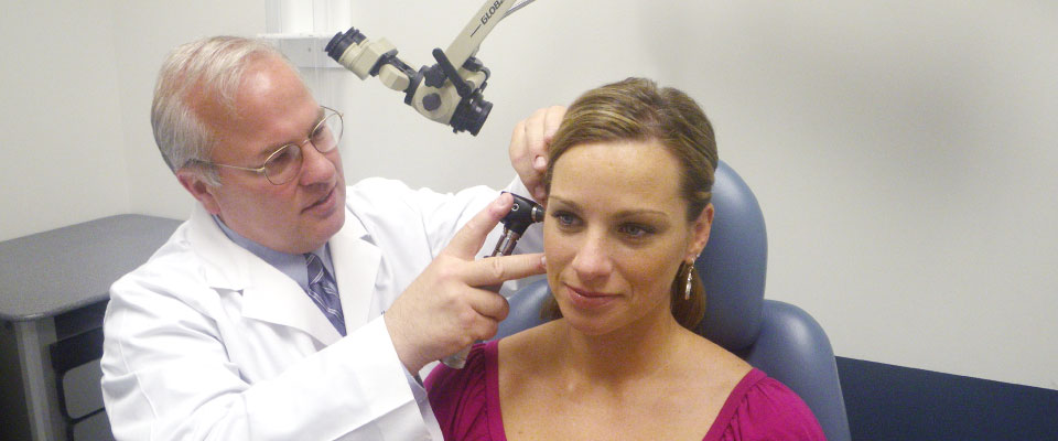
If you are experiencing hearing loss, you should see an ENT (ear, nose, and throat) specialist, or otolaryngologist, who can make a specific diagnosis for you, and talk to you about treatment options, including surgical procedures. A critical part of the evaluation will be a hearing test (audiogram) performed by an audiologist (a professional who tests hearing function) to determine the severity of your loss as well as determine if the hearing loss is conductive, sensorineural, or a mix of both.
Based on the results of your hearing test and what your ENT specialist’s examination shows, as well as results from other potential tests such as imaging your ears with a CT or MRI, the specialist will make various recommendations for treatment options.
The treatment options can include:
- Observation with repeat hearing testing at a subsequent follow up visit
- Evaluation and fitting of a hearing aid(s) and other assistive listening devices
- Preferential seating in class for school children
- Surgery to address the cause of hearing loss
- Surgery to implant a hearing device
These conditions may not, but likely will, need surgery:
- Cholesteatoma
- Bony lesions
- Aural atresia
- Otitis media (if chronic or recurrent)
- Severe retraction of the tympanic membrane
- A hole in the ear drum
- Damage to the middle ear bones
- Otosclerosis
Many types of hearing loss can also be treated with the use of conventional hearing or an implantable hearing device. Again, your ENT specialist and/or audiologist can help you decide which device may work best for you and your lifestyle.
What Questions Should I Ask My Doctor?
- What is the cause of my hearing loss?
- Will my hearing loss likely get worse with time?
- What are my treatment options?
- What are the risks of the surgery you are recommending?
- Do you do this surgery frequently?